Sub Topics: Molecular and Cellular Neuroscience, Cognitive Neuroscience,...
Sub Tracks Clinical Neurology, Neurodegenerative...
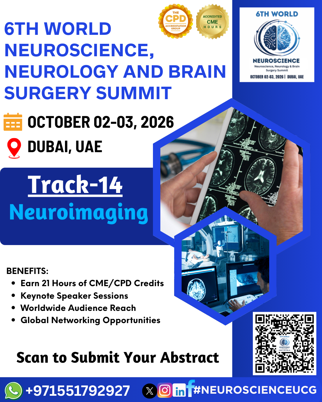
SUB TOPIC; Structural Neuroimaging, Functional
Neuroimaging, Diffusion Imaging,Advanced Neuroimaging Techniques, Pediatric
Neuroimaging, Neuroimaging in Neurodegenerative Diseases,Neuroimaging in
Epilepsy
Neuroimaging refers to the use of various
imaging techniques to visualize the structure, function, and activity of the
brain and nervous system. These techniques allow for the non-invasive
observation of brain anatomy, pathology, and physiological processes, providing
valuable insights for both research and clinical applications. Neuroimaging
plays a crucial role in diagnosing neurological conditions, planning surgeries,
monitoring disease progression, and understanding brain function.
Types of Neuroimaging
Structural Neuroimaging These imaging techniques provide detailed
images of the brain’s anatomy and are primarily used to assess physical
abnormalities or changes in brain structure.
Magnetic Resonance Imaging (MRI):
MRI uses magnetic fields and radio waves to produce
high-resolution images of the brain and spinal cord.
T1-weighted MRI gives clear details of brain anatomy, while
T2-weighted MRI is better for detecting abnormalities like lesions, edema, and
tumors.
Diffusion Tensor Imaging (DTI), a form of MRI, tracks water
molecule movement and assesses the integrity of white matter pathways in the
brain, helping to evaluate diseases like multiple sclerosis and stroke.
Computed Tomography (CT) Scan:
CT uses X-rays to create cross-sectional images of the
brain. It is particularly useful in emergency settings to quickly detect
conditions such as hemorrhages, strokes, or traumatic brain injury.
CT Angiography (CTA) can visualize the brain’s blood vessels
to diagnose conditions like aneurysms or vascular malformations.
Magnetic Resonance Angiography (MRA):
A non-invasive imaging technique used to view blood vessels
in the brain, often used for detecting arterial stenosis, aneurysms, or
arteriovenous malformations (AVMs).
Functional Neuroimaging These methods measure brain activity by
detecting changes in blood flow, metabolic activity, or electrical activity,
providing insights into brain function and how it changes in response to
various stimuli or tasks.
Functional Magnetic Resonance Imaging (fMRI):
FMRI measures brain activity by detecting changes in blood
oxygenation levels (the BOLD signal), which correlate with neural activity.
It is widely used in cognitive neuroscience to study brain
regions involved in tasks such as language, memory, emotion, and motor skills.
fMRI is also employed in pre-surgical mapping of critical
brain regions like those involved in motor or language functions.
Positron Emission Tomography (PET):
PET involves injecting a radioactive tracer into the
bloodstream to assess brain metabolism and blood flow. It is commonly used to
diagnose and study neurodegenerative diseases (e.g., Alzheimer's disease),
epilepsy, and brain tumors.
Amyloid PET scans are especially useful in detecting amyloid
plaques, a hallmark of Alzheimer's disease.
Single-Photon Emission Computed Tomography (SPECT):
SPECT is similar to PET but uses different radioactive
tracers to evaluate blood flow and brain activity.
It is often used in conditions like epilepsy, stroke, and
neurodegenerative diseases.
Diffusion Imaging Diffusion imaging, particularly Diffusion Tensor
Imaging (DTI), provides information about the brain’s white matter pathways,
which are crucial for communication between different brain regions. This
technique is helpful in identifying and characterizing brain injuries,
demyelinating diseases, and disruptions in neural connectivity.
Diffusion Tensor Imaging (DTI):
DTI tracks the movement of water molecules along axonal
fibers, revealing detailed information about the orientation and integrity of
white matter tracts.
DTI is particularly useful in evaluating conditions like
multiple sclerosis, traumatic brain injury, and stroke.
Advanced Neuroimaging Techniques These specialized imaging
techniques provide deeper insights into brain chemistry, metabolism, and more
subtle aspects of brain function.
Magnetic Resonance Spectroscopy (MRS):
MRS is an advanced form of MRI that provides data on the
concentration of various brain metabolites such as N-acetylaspartate (NAA),
choline, and creatine.
It is used to assess metabolic changes in the brain in
conditions like brain tumors, epilepsy, and neurodegenerative disorders.
Arterial Spin Labeling (ASL):
ASL is a type of MRI that measures cerebral blood flow
non-invasively. It is used to assess brain perfusion, helping in the evaluation
of conditions like stroke and vascular dementia.
Quantitative Susceptibility Mapping (QSM):
QSM is a technique used to map the distribution of magnetic
susceptibility in the brain, which can be related to the concentration of
substances like iron and myelin. It is useful in conditions like Parkinson's
disease and multiple sclerosis.
Neuroimaging in Specific Diseases Neuroimaging plays a critical role
in the diagnosis, monitoring, and treatment planning for various neurological
diseases.
Alzheimer’s Disease:
fMRI, Amyloid PET, and MRI help detect early changes such as
hippocampal atrophy and amyloid plaque accumulation.
These imaging techniques are also used to track disease
progression and assess treatment response.
Parkinson’s Disease:
PET and SPECT are used to visualize dopamine activity in the
basal ganglia, aiding in the diagnosis of Parkinsonian syndromes.
MRI is useful for assessing substantia nigra degeneration.
Multiple Sclerosis (MS):
MRI is the gold standard for detecting demyelinating plaques
in the white matter of the brain and spinal cord.
DTI is often used to evaluate disruptions in white matter
integrity in MS patients.
Neuroimaging in Epilepsy Neuroimaging is essential in the evaluation
of epilepsy, helping to identify the epileptogenic focus, assess brain
abnormalities that contribute to seizures, and guide surgical planning.
MRI is commonly used to identify structural abnormalities,
such as hippocampal sclerosis, focal cortical dysplasia, or brain tumors.
fMRI and PET can be used to localize seizure foci and assess
brain activity during seizures.
Neuroimaging in Psychiatry In psychiatry, neuroimaging helps to
understand the underlying brain changes associated with various psychiatric
disorders such as schizophrenia, bipolar disorder, depression, and autism
spectrum disorder (ASD).
fMRI is used to study brain activity and network
connectivity in psychiatric disorders.
PET scans provide insights into neurochemical changes and help visualize areas with altered glucose metabolism or receptor activity.